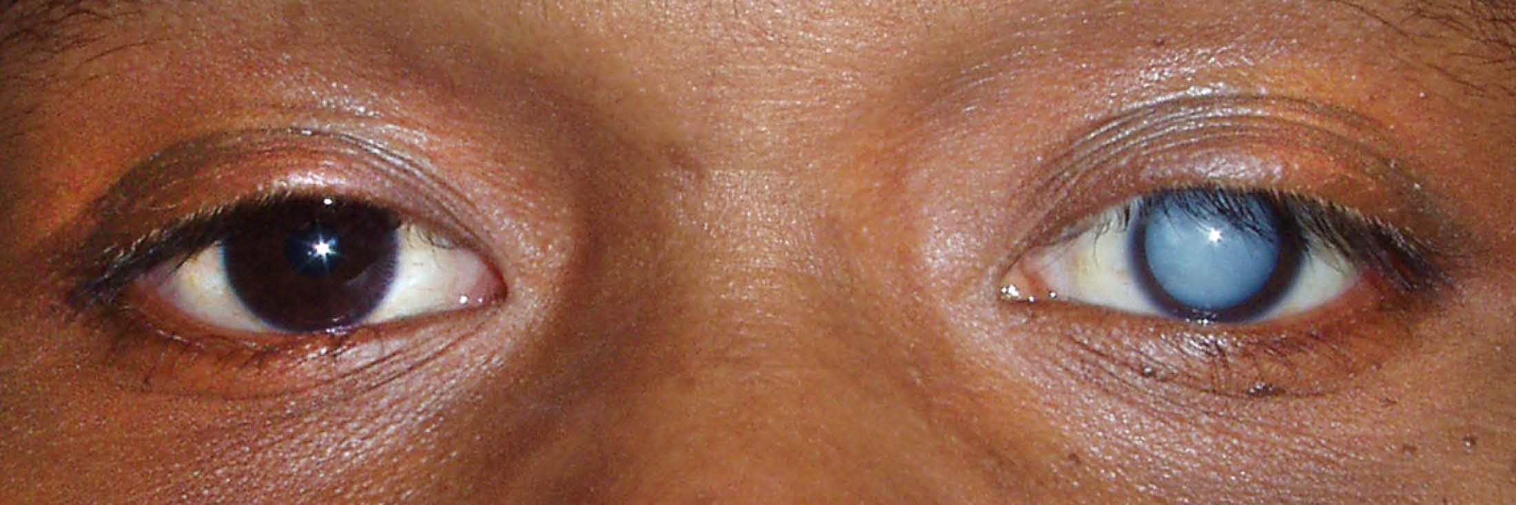
Eye diseases vary widely, but almost all cause visual symptoms. Such diseases can affect any part of the retina, the thin layer of tissue on the inner wall of your eye.
It contains thousands of cones and rods and various nerve cells that coordinate and receive visual information. The retina sends these details directly to your brain via the optic nerve, which allows you to see.
Many rare and common retinal diseases can affect your vision. Such diseases can affect critical eye tissue, causing serious problems and even blindness.
Common types of retinal diseases
Below are some common types of retinal problems:
Floaters:
It usually appears as black, small shapes, visible streaks, spots or squiggles. They seem to hover with the movement of your eyes.
Symptoms:
- Visually, small shapes appear like floating objects, transparent wires and black dots.
- Strings, threads or shapes protrude from the line of sight.
- Floaters are visible when you look at a clear glowing background.
Reasons: Floaters appear when the gel-like substance in the center of the eye becomes extra fluid and clusters and casts a shadow on your retina. They may be:
Age related: Certain retinal conditions such as critical myopia, inflammation at the back of the retina, vitreous hemorrhage, eye hemorrhage, etc. can cause floaters.
Retinal tear:
A retinal tear occurs when the vitreous layer peels off and shrinks from a thin layer of tissue with enough traction to cause damage or a tear in your retina.
Symptoms:
- Instant floaters start in the eye
- Light flashes or photopsia
- In some patients, retinal tears may not show any noticeable symptoms
Reasons:
Starting at birth, the vitreous is attached to your retina. Nevertheless, the gel separates from the retinal lining as a normal aging process. In many people, such a process takes place without any problems. However, people with “sticky” vitreous often experience retinal tears, when the vitreous layer separates from the retina.
Retinal detachment
In this, the retinal layer detaches from the back of your eye, and this happens when the vitreous passes through the retinal tear. This separates the retina from its primary position.
symptoms
Although this is a painless condition, below are some of the symptoms that always occur before retinal detachment:
- Immediate occurrence of floaters within the field of vision
- Light shines in the pompous eye
- Peripheral vision is reduced
- blurred vision
- A shadow on the field of vision
Reasons
- Age-related variation in the eye
- Eye injuries
- Myopia/myopia
- Lattice degeneracy
- Previous cataract surgeries
- Retinal detachment or genetic problem
Also Read: Types of Cataract Eye Treatment with their Cost in India
Diabetic retinopathy
Diabetic retinopathy is caused by an increase in blood sugar levels, which affects the delicate retinal blood vessels.
symptoms
Diabetic retinopathy, in its early stages, is difficult to notice and painless. You won’t be able to notice its symptoms until it is highly magnified. As the condition progresses, it may show the following signs:
- Finally the vision is fading
- Floaters
- Immediate vision loss
- Damaged color vision
- Blurred vision or blank vision areas
Reasons:
In diabetic retinopathy, abnormal enlargement of blood vessels through the retina causes bleeding and scarring. Blood vessels collapse and leak fluid into and down the retina. It causes swelling of the retina, which gradually distorts and blurs vision.
Epiretinal membrane:
An epiretinal membrane is a clear, delicate, thin membrane or scar that appears above the macula. It pulls on the retina and blurs vision.
Symptoms:
It does not cause total blindness, however, it can affect your primary vision. Peripheral vision of the damaged eye remains unchanged.
- Blurred vision
- A critical part of distortion of vision.
Reasons:
It is caused by the formation of fine and thin sheets of fibrous tissue on the macula. Such a membrane acts as a film, obstructing vision. It can contract like scar tissue and pull on your retina, causing macular puckering. All these things lead to swelling of the retina or distorted vision. Seedi Eye Care Centre is one of the most recognized eye care center in Bangalore. Our eye experts help you with retinal diseases and treatments like Cataract, Glaucoma, lasik, Ophthalmology, Refractive Eye Surgery and many more.
Also Read: How Much does Cataract Eye Surgery Cost in Bangalore?
What is the primary function of the retina?
The retina is a thin layer of tissue that contains millions of light-sensitive cells (rods and cones) and is located at the back of the eye. The retina’s primary function is to receive, organize, and send visual information to your brain via the optic nerve. This entire process allows you to see.
What is retinal disease?
Retinal diseases can damage any part of the retina. Untreated retinal diseases can lead to severe vision loss and blindness. With early retina symptoms detection, some retinal diseases can be treated at early stages, while others can be controlled or slowed, or vision can be restored.
Symptoms of retinal disease
Many retinal diseases and conditions present with similar signs and symptoms, including:
- Seeing floaters or flashes of light
- Blurred or distorted vision
- Blind spots in central vision
- Reduced vision
Who is at risk of developing retinal disease?
Certain factors can increase your risk of developing retinal disease, including:
- age
- obesity
- diabetes
- smoking
- Eye trauma
- Family history
If you experience symptoms of retinal disease, consult an eye doctor or expert eye surgeon to diagnose and manage your condition.
Retinitis pigmentosa
Retinitis pigmentosa (RP) is a rare, genetic, eye disease that causes retinal damage and vision loss.
RP is one of the most frequent forms of inherited retinal degeneration.
RP affects about one in 4,000 people worldwide.
In its early stages, retinal cells begin to degenerate in the part of the retina responsible for central-external vision—which can lead to reduced night vision (nyctalopia), loss of central-external visual field, and difficulty seeing in low light. The rapid progression of the disease continues to destroy cells in the central visual field—resulting in tunnel vision, reduced visual acuity, and loss of color vision.
Progression of RP causes symptoms at a similar rate in both eyes.
In its later stages, RP causes sensitivity to bright lights due to intense glare (photophobia) and the appearance of flashing, flashing or rotating lights in the visual field (photopsia).
RP can be caused by several gene mutations, and its progression can vary from person to person—in some cases, central vision is not affected until the person reaches age 50, while in other cases, people experience significant vision loss in early adulthood.
Eventually, most individuals with RP lose a significant amount of their vision.
There is currently no cure for RP. Many patients learn to use low vision devices aimed at enhancing existing central vision to expand the field of vision and eliminate glare.
Central venous occlusion
Central retinal vein occlusion (CRVO) is a condition that occurs when the central vein responsible for draining blood from the retina is blocked – usually as a result of a blood clot. This condition can also be caused by diabetes or glaucoma.
Central retinal vein occlusion usually occurs in only one eye.
There are two different forms of CRVO:
Non-ischemic CRVO- This is a milder form that results in leakage of retinal cells and development of macular edema. This form is usually asymptomatic, although patients may complain of blurred or distorted vision. This type can go into a more severe form.
Ischemic CRVO—a more severe form that causes retinal cells to shut down—results in pain, redness, irritation, and vision loss, with little chance of vision recovery.
Central retinal vein occlusion is usually treated with anti-VEGF injections, which are used to reduce new abnormal blood vessel growth and retinal swelling. Anti-VEGF medications include bevacizumab (Avastin®), ranibizumab (Lucentis®), and aflibercept (Ilia®).
Laser surgery may also be recommended for a more permanent cure.
Branch retinal vein occlusion
Branch retinal vein occlusion (BRVO) occurs when there is an obstruction in a branch of the retinal vein, as opposed to CRVO caused by an obstruction in the central retinal vein. Branch retinal vein occlusion usually occurs as a result of a blood clot blocking blood drainage.
Branch retinal vein occlusion usually causes sudden loss of vision. However, if BRVO does not affect the center of the retina, BRVO may go unnoticed.
Branch retinal vein occlusion is usually treated with anti-VEGF injections and/or laser surgery.
This is a serious complication that can occur as a result of BRVO. This condition causes ischemia, or inadequate blood supply to the retina—resulting in the growth of abnormal new blood vessels. These abnormal blood cells can cause vitreous hemorrhage and further vision loss.